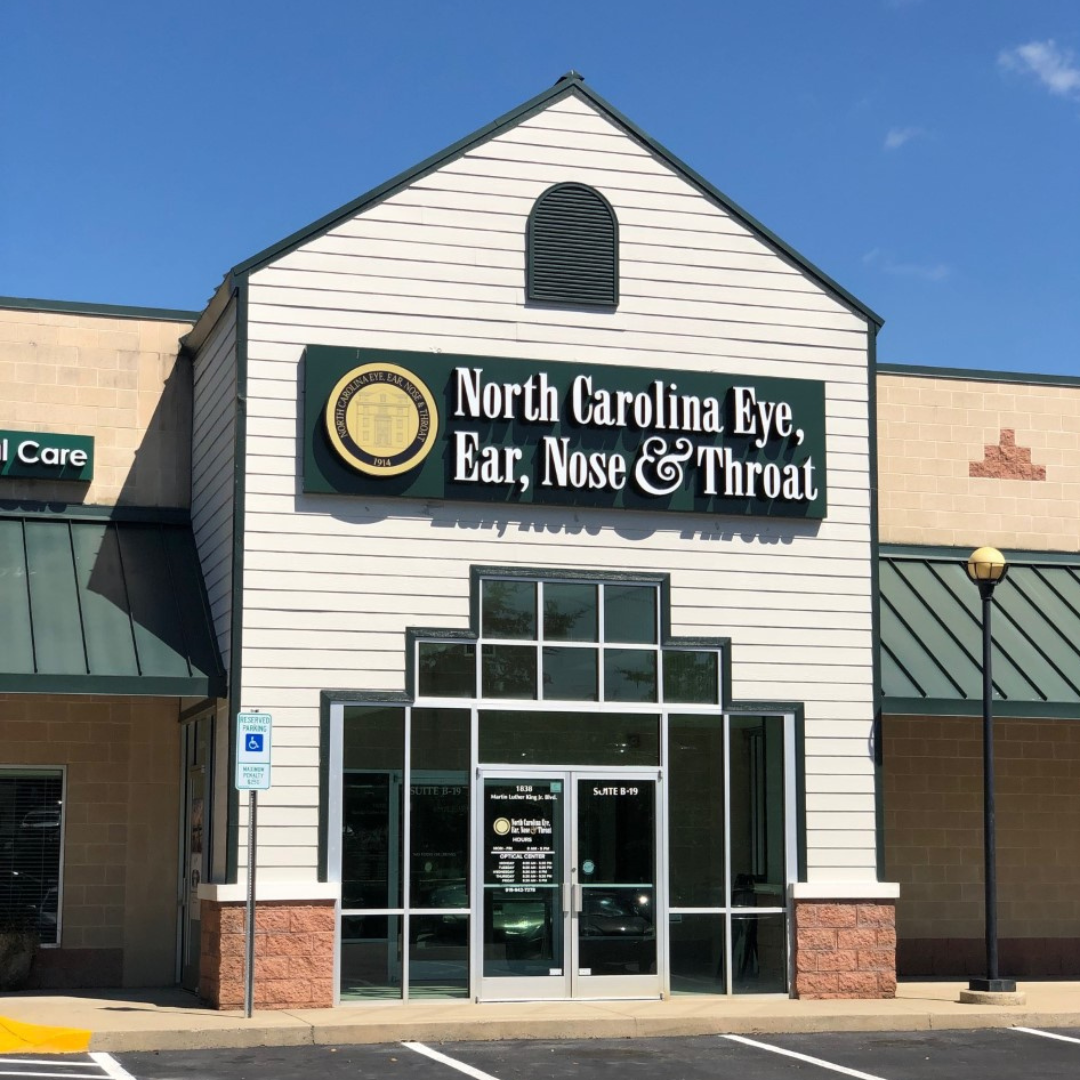
Carolina Ophthalmology Associates Partners with North Carolina Eye, Ear, Nose & Throat-Chapel Hill and aligns with Duke Health
Carolina Ophthalmology Associates Partners with North Carolina Eye, Ear, Nose & Throat-Chapel Hill and aligns with Duke HealthDURHAM, N.C. – Beginning Jan. 1, 2024, Read More
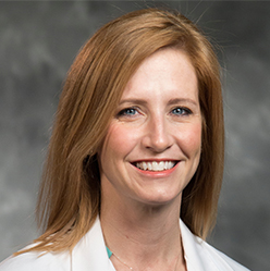
Dr Sara Grace Presents Key Opthalmology Takeaways Related to Dermatology
North Carolina Eye, Ear, Nose & Throat Pediatric Ophthalmologist, Dr. Sara Grace, presented key ophthalmology takeaways related to dermatology in her session “Seeing Eye Read More
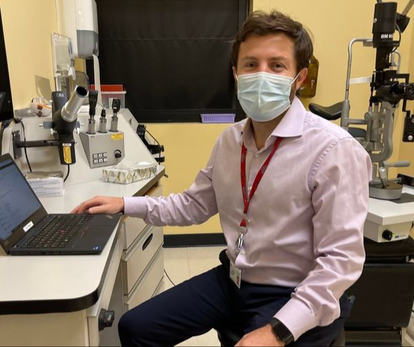
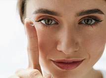
Eye Conditions, including Glaucoma, Cataract Surgery
North Carolina Eye, Ear, Nose & Throat is proud to welcome Alexander S. Barsam, MD. The comprehensive ophthalmologist treats various eye conditions, including glaucomaand Read More
Hearing Tests for Adults - Dr. Shannon Bornstein
Dr. Shannon Bomstein. She’s a clinical audiologist who offers hearing aid fittings and hearing tests for adults. Dr. Bomstein sees patients in our North Read More
Hearing Aid Fittings, Hearing Tests by Dr. Schyler House
Audiology: Cary, DurhamDr. Schyler House. She’s a clinical audiologist who offers hearing aid fittings and hearing tests for adults. Dr. House sees patients in our Read More